The E protein: A small but mysterious structure

July 10, 2020
Luise Kandler

Introduction

The novel Coronavirus SARS-CoV-2 incorporates various structural proteins in its protective coat. In order to find a potential drug target against the spreading pandemic, a lot of scientific research focusses on the characteristic spike glycoprotein as a therapeutic target. But apart from the spikes, several other structural proteins were found to decorate the virus hull of which the envelope protein (E protein) is the smallest one, consisting of only 75 amino acids. Even though it is an integral membrane protein, the envelope protein is also localized in the host ER, Golgi, and ERGIC (ER-Golgi intermediate compartment) [1], where it is essential for virus formation.

Interestingly, research on this protein can shed light on the origin of the novel coronavirus, which currently dominates everyday life all over the globe. Sequence comparisons of several different envelope proteins strengthen the assumption that SARS-CoV-2 may originate from Bat-CoV or Pangolin-CoV due to a high sequence homology [2]. The E protein of SARS-CoV-2's "older brother" SARS-CoV exhibits a nearly identical sequence with 91% homology [2] as well and has been structurally determined based on nuclear magnetic resonance (NMR) data. Yet, until now, solving the 3D structure of SARS-CoV-2 E protein turns out to be quite challenging, and hence no experimental structure is available for the new coronavirus [3].

Structure comparison with SARS-CoV E protein

We, as structural biologists aim to uncover and refine the structures of as many of the novel virus's proteins as possible. But, as long as no structures of SARS-CoV-2 E protein have been solved, this could only be achieved by comparing it to the existing structures of SARS-CoV envelope protein.

Topology and structural features

The topology of SARS-CoV E protein is mainly separated into three domains:  A short hydrophilic N-terminus, that has an identical sequence in SARS-CoV-2 [3] and works as a Golgi-targeting signal; a long mainly hydrophobic transmembrane domain (TMD), and a long hydrophilic C-terminal domain. Studies on the question whether the C- and the N-terminus are luminal or cytoplasmic have had different results, suggesting that the E protein’s topology could differ depending on its multiple functions [3].
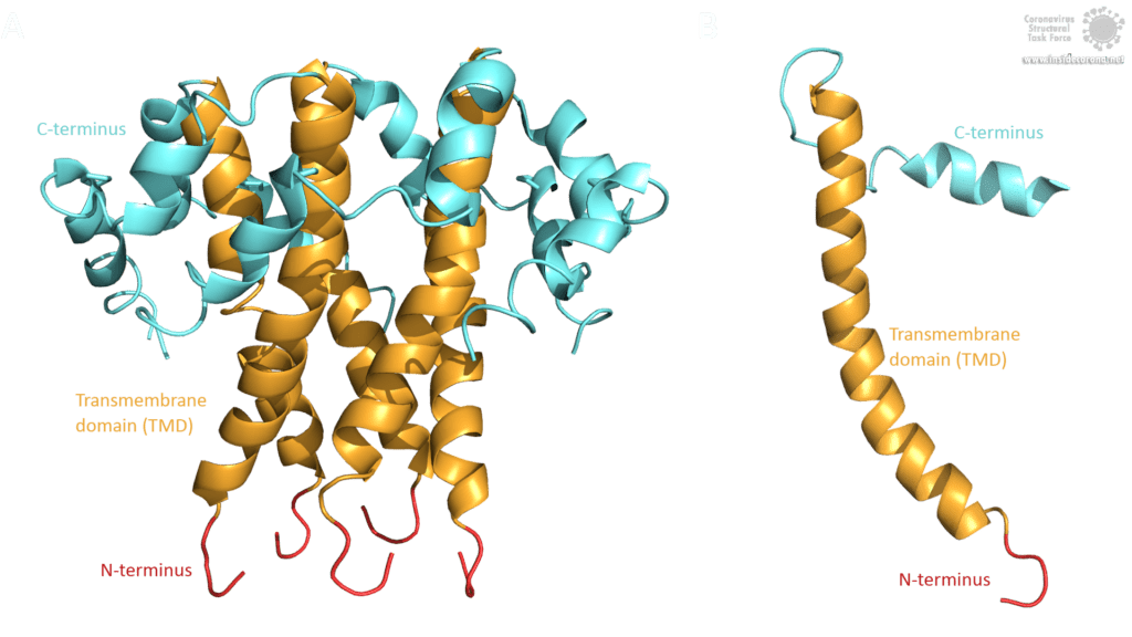
Domains of the E protein in SARS-CoV
Figure 1: The topology of the SARS-CoV E protein is colored to indicate the different parts. The N-terminus is displayed in red, the Transmembrane domain (TMD) in bright orange and the C-terminus in cyan. (A) The topology of E as an oligomer. (B) The topology of E as a monomer.
Image by Luise Kandler

The E protein of SARS-CoV comprises several interesting structural features: A long α-helix with amphipathic parts forms the Transmembrane domain (TMD). The C-terminus, however, incorporates a short α-helix which is believed to be in a dynamic equilibrium with a less abundant β-coil-β-motif. Both helices are connected by a turn [4]. The β-coil-β-motif with a conserved proline residue (Pro-54) has been proposed to function as Golgi targeting signal, and to switch its conformation in order to alter the E protein's function in the host cell [4]. Furthermore, the C-terminus contains a PDZ-binding motif (PBM) at residues 73-76 [3]. This PBM domain slightly differs in coronaviruses but a DLLV motif is conserved in the E proteins of  SARS-CoV, Bat-CoV, and SARS-CoV-2 [2]. Unfortunately, there are no PDB structures available that exhibit the β-coil-β-motif nor the PBM domain.
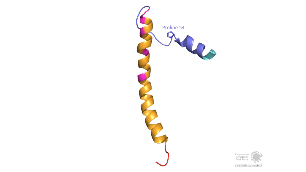
Structural features of E protein monomer.
Figure 2: Front view of the structural features of SARS-CoV E protein. The hydrophilic residues of the amphipathic α-helix are displayed in magenta, the hydrophobic rest in bright orange. The short C-terminal α-helix is colored in cyan and slate blue. Slate blue indicates residues that are in dynamic equilibrium with the β-coil-β-motif.
Image by Luise Kandler

Structural variants - Oligomerization and posttranslational modifications

The E protein comes in two different forms. Apart from a monomeric structure, the protein also oligomerizes to form a pentameric viroporin in the host cell's Golgi membrane. Whether the E proteins that are embedded in the viral hull are pentamers or monomers is not yet clear. Oligomerization is induced by the amphipathic α-helix of the TMD [3] and is proposed to be mainly mediated by residue Val-25 as well as residue Asn-15 being slightly involved [3]. Both residues are conserved in SARS-CoV-2 as well. To anchor the pore in the Golgi membrane, the hydrophobic amino acids of the TMD orientate towards the phospholipids. Additionally, basic positively charged residues interfere with the negatively charged phospholipids via electrostatic interactions [3].
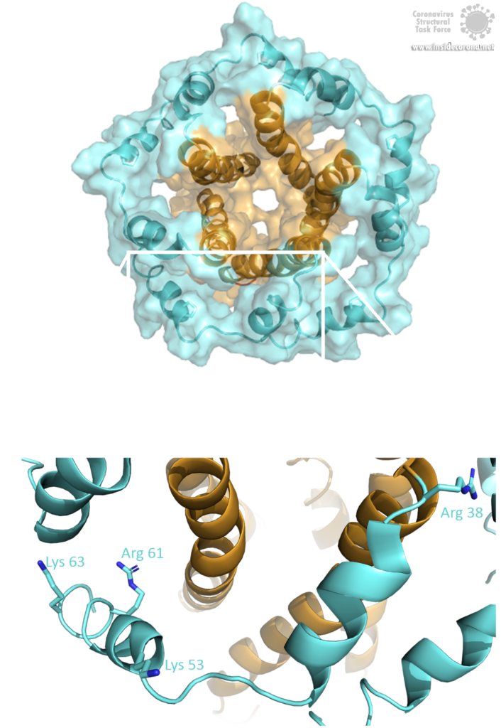
Top view of Ion channel pore and positively charged residues of the C-terminus.
Figure 3: (A) Top view of the SARS-CoV viroporin surface displaying the ion channel built up in the center. (B) Positively charged residues, which can interfere with the negatively charged membrane lipids to anchor the pore.
Image by Luise Kandler

Other structural variants are obtained by posttranslational modifications, which have been detected in the E protein of SARS-CoV and other coronaviruses. Palmitoylation is the addition of palmitic fatty acid to cysteine residues which increases the protein's hydrophobicity. Hence, the palmitoylation of E protein assists in membrane anchoring and probably aids Golgi targeting. Ubiquitination of the E protein might function as negative regulation of E protein levels [3]. It has been shown that the optimal amount of E protein present in the host cell is important for a successful production of new viruses. Another modification, namely glycosylation, adds oligosaccharide fragments to asparagine residues in a certain motif (Asn-X-Ser/Thr) which is also conserved in E protein. In SARS-CoV, residue Asn-66 embedded in the motif Asn-Ser-Ser was proven to be glycosylated. This may help to recruit chaperone proteins of the host cell to aid in the correct folding of newly synthesized viral proteins as well as in defense against the host immune system. Experimental data suggest that glycosylation of Asn-66 might also promote E protein's monomeric functions as it prevents oligomerization [3].

Connecting function and structure

To understand a molecule's biological function is the main goal of experimental structure determination. A viral protein can be targeted by drugs best if the atomic structure is known. The envelope protein has various structural conformations and thus multiple functions, both as a monomer and as a pentamer.

Monomeric functions: Golgi-targeting and viral assembly

The E protein comprises a Golgi-targeting signal in the β-coil-β motif of the C-terminus and another one in the N-terminal domain. Additionally, palmitoylation is believed to be involved in this function. Accordingly, after being translated at the ER, the E protein is located to the Golgi membrane. From there, the virus acquires the membrane for a new viral envelope [3]. Once the protein is located to the Golgi, one of its main functions as a monomer is in viral assembly, which means the process of gathering all the viral macromolecules (proteins and the RNA genome) to form a virus-like particle. During this assembly, the virus-like particle buds into the lumen of ERGIC and follows the way through the host cell's secretory pathway. Several experiments confirm the involvement of the envelope protein together with the membrane protein (M) into this process. It has been proposed that the E protein rather induces membrane curvature and scission, whereas the M protein may coordinate viral assembly. Nevertheless, SARS-CoV-infected cells still produce virus-like particles in the absence of E protein, but virus trafficking to the cell surface and viral secretion are hampered, resulting in a lower number of mature virions, an atypic morphology and a higher rate of propagation incompetent virions [3]. Further investigation will be necessary to analyze the exact mechanism behind the membrane formation of virions.
After finding its way through the secretory pathway, the mature virion is released from the host cell. The process of detaching from the host membrane is known as scission and is either coordinated by the virus's own scission proteins or by the host cell's scission machinery (called ESCRT). Which one is the case for SARS-CoV-2 is still unclear. Infected cells lacking the scission machinery exhibit a “beads-on-a-string” morphology, with the virions being stuck to the host membrane in an elongated shape. This morphology was found in influenza-infected cells lacking the M2 protein, which proves that M2 is involved in this scission process. Given that SARS-CoV E protein is suggested to be functionally equivalent to M2, due to similar structural features, the E protein is proposed to be involved in the scission process as well [3].

Sars-CoV-2 life cycle.
Figure 4: (A) The Lifecycle of Sars-CoV-2. The envelope protein is colored in yellow. After its translation at the ER (5) the E protein is transported to the Golgi, where it is involved in viral assembly (6) and the release of the virus (7). (B) Zoomed-in image of the E protein’s involvement in the lifecycle.
Image by Ann (Hui) Liu, https://animationlab.utah.edu/ [5]

Pentameric functions: Ion-channel activity

While located at the Golgi, some of the SARS-CoV E proteins oligomerize and form a pentameric viroporin. These pores of SARS-CoV E protein function as ion channels. They mainly favor the transport of Na+ and K+, but were also found to be permeable for Ca2+ ions and eventually for H+ ions. Even though the primary purpose of transporting cations is not yet clear, Ca2+ is proposed to trigger the inflammatory response seen in acute respiratory distress syndrome [7].
Residue Asn-15 has been suggested to act as a "filter" for this ion selectivity [6], which can further be affected by the charge of the membrane's lipid head group. Deletion of the envelope protein in its pentameric form demonstrates that ion channel activity is not essential for viral replication, but yet attenuates the virulence [8].
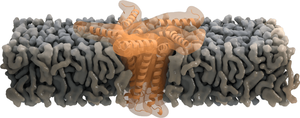
This illustration shows the E protein as a pentameric ion channel embedded in a membrane.
Figure 5: Illustration of the E protein ion channel anchored in a membrane.
Image by Thomas Splettstoesser, www.scistyle.com

Pathogenesis and the E protein as a potential drug target

Interactions of viral proteins with host cell proteins de-regulate many physiological processes. In patients suffering from SARS-CoV infections, these de-regulating protein-protein interactions greatly contribute to pathogenesis. Some of the observed symptoms are also present in a SARS-CoV-2 infected patient.
Interactions of the envelope protein with proteins of the host cell are mediated by its PDZ-binding motif (PBM) at the very end of the C-terminus. The motif binds to the PDZ domain of adaptor proteins, which are subsequently bound by other cellular proteins, activating a signaling cascade that may result in pathogenesis. Some of these interactions were proposed or even proven to induce symptoms like lymphopenia [9], changes in fluid volume, blood pressure, and water homeostasis, as well as tissue damage, edema and acute respiratory distress syndrome (ARDS) [10], due to an overexpression of inflammatory cytokines (which are also regulated by the leader protein nsp1). Another protein-protein interaction was found to disrupt tight junctions of pulmonary epithelial cells in the lungs. This eventually results in an epithelial barrier failure and virions breaking through the alveolar wall causing a systemic infection [11]. O. Wittekindt writes [12]: "The breakdown of the epithelial barrier is a hallmark in respiratory distress syndromes (...)" Furthermore, the ion channel activity of the E protein activates the inflammatory pathway by channeling Ca2+ resulting in lung damage in infected mice [7]. Inhibition of the viroporin by hexamethylene amiloride (HMA) [8] reduces the activation of the inflammasome, which makes the ion channel of E protein a potential therapeutic target. Additionally, as a part of the host cell's viral defense, the ER stress response is activated, once the protein folding capacity of the ER is overloaded by additional expression of viral proteins. This can lead to apoptosis of the host cell. However, experiments confirm that the E protein contributes to pathogenesis by suppressing the ER stress response to maintain the survival of the host cell [3].
As a potential target for drug treatment, protein-protein interactions of the E protein are quite promising. Its PBM domain can bind cellular proteins that are involved in pathogenesis. Experimental truncation of this domain shows that it may be possible to find a live vaccine with a mutated but intact PBM and thus attenuated pathogenicity. Identifying more interacting partners could provide a more targeted therapy, though. The absence of E protein furthermore leads to reduced viral titers, crippled viral maturation, and propagation-defective progeny [3], making E protein-deficient virions also a potential vaccine candidate.

In conclusion, one could say that the E protein of SARS-CoV-2 is another valuable drug target. While the protein's "older brother" SARS-CoV E protein gives us much insight into its function, an experimental structure determination of SARS-CoV-2 E protein would be highly desirable. Until then, the envelope protein SARS-CoV-2 remains a small but mysterious structure.

Best PDB structures available

  1. 2MM4: This NMR structure is a monomer of SARS-CoV envelope protein (E). It covers the transmembrane domain completely and the C- and N-terminus partly. The structure is involved in membrane curvature, membrane scission, and viral assembly.
  2. 5X29: This NMR structure is an oligomer of SARS-CoV envelope protein (E). The structure is a pentamer of five identical monomers. It covers the transmembrane domain completely and the C- and N-terminus partly. This structure functions as a membrane-anchored ion channel.

Literature

[1] J. Nieto-Torres, M. DeDiego, E. Álvarez, J. Jiménez-Guardeño, J. Regla-Nava, M. Llorente, et al.: Subcellular location and topology of severe acute respiratory syndrome coronavirus envelope protein, Virology, 2011

[2] M. Bianchi, D. Benvenuto, M. Giovanetti, S. Angeletti, M. Ciccozzi, S. Pascarella: Sars-CoV-2 Envelope and Membrane proteins: differences from closely related proteins linked to cross-species transmission, Preprint, 2020

[3] D. Schoeman, B. Fielding: Coronavirus envelope protein: current knowledge, Virology Journal, 2019

[4] Y. Li, W. Surya, S. Claudine, J. Torres: Structure of a Conserved Golgi Complex-targeting Signal in Coronavirus Envelope Proteins, The Journal Of Biological Chemistry, 2014

[5] Ann (Hui) Liu, in https://animationlab.utah.edu/

[6] K. Pervushin, E. Tan, K. Parthasarathy, X. Lin, F. Jiang, D. Yu, A. Vararattanavech, T. Soong, D. Liu, J. Torres: Structure and Inhibition of the SARS Coronavirus Envelope Protein Ion Channel, PloS Pathogens, 2009

[7] J. Nieto-Torres, C. Verdiá-Báguena, J. Jimenez-Guardeño, J. Regla-Nava, C. Castaño-Rodriguez, R. Fernandez-Delgado, et al.: Severe acute respiratory syndrome coronavirus E protein transports calcium ions and activates the NLRP3 inflammasome, Virology, 2015

[8] J. Nieto-Torres, M. DeDiego, C. Verdiá-Báguena, J. Jimenez-Guardeño, J. Regla-Nava, R. Fernandez-Delgado, et al.: Severe acute respiratory syndrome coronavirus envelope protein ion channel activity promotes virus fitness and pathogenesis, PLoS Pathogens, 2014

[9] Y. Yang, Z. Xiong, S. Zhang, Y. Yan, J. Nguyen, B. Ng, et al.: Bcl-xL inhibits T-cell apoptosis induced by expression of SARS coronavirus E protein in the absence of growth factors, Biochemical Journal, 2005

[10] J. Jimenez-Guardeño, J. Nieto-Torres, M. DeDiego, J. Regla-Nava, R. Fernandez-Delgado, C. Castaño-Rodriguez, et al.: The PDZ-binding motif of severe acute respiratory syndrome coronavirus envelope protein is a determinant of viral pathogenesis, PLoS Pathogens, 2014

[11] K. Teoh, Y. Siu, W. Chan, M. Schlüter, C. Liu, J. Peiris, et al.: The SARS coronavirus E protein interacts with PALS1 and alters tight junction formation and epithelial morphogenesis, Mol Biol Cell, 2010

[12] O. Wittekindt: Tight junctions in pulmonary epithelia during lung inflammation, Springer Verlag, 2016

Corinna the Corona Cactus

@
Corinna works as an outreach person for all plant-related business and as a mascot. She gathered previous experience in the garden center, and even though she can be a bit spiky, she likes to cuddle and lie in the sun.
More about this author

Helen Ginn

Senior Research Scientist @ Diamond Light Source, Oxfordshire, UK
Dr Helen Ginn is a senior research scientist at Diamond Light Source in the UK and a computational methods developer in structural biology. She is currently working on Representation of Protein Entities (RoPE) for structural biologists to interpret subtle conformational changes in dynamic protein systems. She has developed Vagabond for torsion angle-driven model refinement and […]
More about this author

Nick Pearce

Assistant Professor @ SciLifeLab DDLS Fellow
Nick obtained his undergraduate degree in Physics from the University of Oxford in 2012, and then his PhD in Systems Approaches to Biomedical Sciences in 2016. He moved to Utrecht in the Netherlands in 2017 to work with Piet Gros, where he obtained an EMBO long-term fellowship and worked on analysing disorder in macromolecular structures. […]
More about this author

Mathias Schmidt

Molecular Life Sciences M.Sc. Student @ Hamburg University
Mathias is currently doing his Master's degree in Molecular Life Sciences at the University of Hamburg and has been an auxiliary scientist in the Corona Structural Taskforce since March 2022. There he is working on the question of the origin of SARS-CoV-2. His undergraduate research focuses on the development of synthetic molecular mechanisms to regulate […]
More about this author

David Briggs

Principal Laboratory Research Scientist @ Francis Crick Institute in London, UK
David Briggs is a Principal Laboratory Research Scientist in the Signalling and Structural Biology lab at the Francis Crick Institute in London, UK. A crystallographer by training, his work focuses on the biophysical and structural characterisation of human extracellular proteins involved in the synapse, which have important ramifications in both psychiatric and neurodegenerative disorders. He […]
More about this author

Lisa Schmidt

Web Developer and Illustrator @ Mullana
Lisa Schmidt is a freelance illustrator who studied Multimedia and Communication (BA) in Ansbach, Germany. Her work is focused on visualising topics around science and technology. She joined the Coronavirus Structural Task Force as media designer, where she does web design, 3D rendering for scientific illustrations and outreach work.
More about this author

Philip Wehling

Nanosciences M.Sc. Student @ Institute for Nanostructure and Solid-State Physics, Hamburg University
Philip has long had an enthusiasm for biological processes which is paired with an analytical understanding of the world. After having worked for a long time as a registered nurse in various fields, he first studied mathematics and finally nanosciences. During a lecture series in preparation for a bachelor's thesis, he came into contact with […]
More about this author

Binisha Karki

Postdoctoral Research Associate @ BioNTech SE
Binisha works as a research associate at BioNTech where she works on the development of COVID-19 vaccine and cancer immunotherapies. She graduated as a Molecular Biology major from Southeastern Louisiana University in May 2019. Post-graduation she worked as a research technician in the Chodera Lab performing biophysical measurements of model protein-ligand systems for computational chemistry […]
More about this author

Binisha Karki

Wissenschaftliche Mitarbeiterin @ BioNTech SE
Binisha ist als wissenschaftliche Mitarbeiterin bei BioNTech angestellt und arbeitet an der Entwicklung von Impfstoffen gegen COVID-19 sowie Krebsimmuntherapien. Sie beendete ihr Studium der Molekularbiologie an der Southeastern Louisiana University im Mai 2019. Anschließend arbeitete sie als Forschungstechnikerin im Chodera-Lab, wo sie biophysikalische Messungen an Modellen von Protein-Liganden-Systeme für computerchemische Benchmarks durchführte.
More about this author

Hauke Hillen

Assistant Professor at the University Medical Center Göttingen & Group Leader at the MPI for Biophysical Chemistry @ University Medical Center Göttingen
Hauke ist Biochemiker und Strukturbiologe. Mit seinem Forschungsteam untersucht er mittels Röntgenkristallografie und Kryo-Elektronenmikroskopie die Struktur und Funktion von molekularen Maschinen, die für die Genexpression in eukaryotischen Zellen verantwortlich sind. Er interessiert sich dabei besonders dafür wie genetisches Material außerhalb des Zellkerns exprimiert wird, zum Beispiel in menschlichen Mitochondrien oder durch Viren im Zytoplasma.
More about this author

Richardson Lab

Richardson Lab @ Duke University, Durham, North Carolina, USA
The long-term goal of the Richardson lab is to contribute to a deeper understanding of the 3D structures of proteins and RNA, including their description, determinants, folding, evolution, and control. Their approaches include structural bioinformatics, macromolecular crystallography, molecular graphics, analysis of structures, and methods development, currently focussed on the improvement of structural accuracy. In this […]
More about this author

Holger Theymann

Agile Leadership Coach @ mehr-Freu.de GmbH
Holger keeps websites running. He makes data from scientific databases appear in nice tables. He also has an eye on keeping the sites fast, safe and reliable. His experience as a software developer, systems architect, agile project manager and coach enabled the Task Force to get the whole process well organized and he even taught […]
More about this author

Florens Fischer

Biology M.Sc. Student @ Rudolf Virchow Center, Würzburg University
Florens is studying biology (M.Sc.) and worked in the Task Force as a student assistant. He has focused on bioinformatics and supports the work on automation of scripts and structuralization of big data with machine learning. He also supported the team in other areas, such as scientific research.
More about this author

Ezika Joshua Onyeka

Public Health M.Sc. student @ Hamburg University of Applied Sciences
Joshua joined Thorn Lab as a student assistant. He is a Public Health practitioner, holds a bachelor's degree in Public Health and is currently enrolled at Hamburg University of Applied Sciences for his MPH. He has helped in implementing some vaccination programmes to improve immunisation coverage and training of immunisation frontline health workers. For the […]
More about this author

Katharina Hoffmann

Molecular Biology M.Sc. student @ Institut für Nanostruktur und Festkörperphysik, Universität Hamburg
Katharina worked as a student assistant at Thorn Lab. Normally, she studies molecular biology at the University of Hamburg. In her master's thesis, which was put on hold by Corona, she is working on the interruption of bacterial communication. Since the lockdown, she has been digging around in databases and analyzing sequences. She never thought […]
More about this author

Nicole Dörfel

Media Designer @
Nicole Dörfel ensures that we and our work are looking good! She is the illustrator, media designer and the artistic soul of the Task Force. She works her magic both in print and digitally—her focus is general media design. In the Task Force, she is mainly responsible for graphics, photo editing, design of all our […]
More about this author

Pairoh Seeliger

Administration Assistant @ Institute for Nanostructure and Solid-State Physics, Hamburg University
Pairoh Seeliger is the admin wizard of the Task Force. She takes care of media requests, handles any logistical issues that come up and makes sure our science doesn’t sound too complicated in our German outreach efforts. She self-describes as "a jack of all trades with a University education in German studies and business administration, […]
More about this author

Oliver Kippes

Biochemistry B.Sc. Student @ Rudolf Virchow Center, Würzburg University
Oli is studying biochemistry (B.Sc) and has completed a training as an IT specialist prior to his studies. With the combined knowledge of his studies and training, he helps maintaining the structural database, programs applications for it and supports the team in literature research. In spite of his study, structural biology was still a new […]
More about this author

Luise Kandler

Biochemistry B.Sc. Student @ Rudolf-Virchow Center, Würzburg University
Luise is a B.Sc. student in biochemistry at the University of Würzburg and joined the Task Force during the first Corona lockdown. She did her bachelor's thesis with the Thorn Lab, where she learned programming with Python and worked on the implementation of a GUI for our machine learning tool HARUSPEX in Coot. In the […]
More about this author

Ferdinand Kirsten

Biochemistry B.Sc. Student @ Rudolf Virchow Center, Würzburg University
Ferdinand did his bachelor's thesis at Thorn Lab on solvent exchange and interactions in macromolecular crystallography. Still new to the world of crystallography and structural refinement, he tries to help wherever he can, with a main focus on literature and genome research as well as structural refinement with Coot. Even if he's more of the […]
More about this author

Kristopher Nolte

Biochemistry B.Sc. Student @ Rudolf-Virchow Center, Würzburg University
Kristopher joined Thorn Lab as part of his bachelor thesis. In this thesis he refined aspects of the diagnostic tool for graphical X-Ray data analysis (AUSPEX) with the help of machine learning. But since the corona crisis halted all our lives, he contributes to the Task Force by using his knowledge of bioinformatics and programming […]
More about this author

Erik Nebelung

Nanoscience M.Sc. Student @ Institute for Nanostructure and Solid-State Physics, Hamburg University
Erik is studying nanoscience with a focus on biochemical methods and applications. From August 2020 till January 2021 he pursued his studies at the iNano institute in Aarhus, before starting his master's thesis back in Hamburg. He had his first taste of protein crystallization during his bachelor's thesis work and this sparked his interest in […]
More about this author

Toyin Akinselure

Nanoscience M.Sc. Student @ Institute for Nanostructure and Solid-State Physics, Hamburg University
Toyin ist a microbiologist and presently an M.Sc. student in nanoscience with a focus on nanobiology and nanochemistry. She is interested in scientific research especially in protein chemistry and drug discovery. In the previous autumn and winter, she interned with two research projects, one in drug discovery and the other in protein structure. She found […]
More about this author

Lea von Soosten

Physics M.Sc. Student @ Institute for Nanostructure and Solid-State Physics, Hamburg University
Lea is a M.Sc. physics student with a great interest in everything related to biology. Even though she comes from a different field, she joined the team to expand her knowledge in biochemistry and help the Task Force with a main focus on literature research. Also, she loves drawing!
More about this author

Sabrina Stäb

Biotechnology M.Sc. Student @ Institute for Nanostructure and Solid-State Physics, Hamburg University
Sabrina is studying biochemistry (M.Sc.) and works as a research assistant for the Thorn Lab and the CSTF. During her bachelor thesis on "Crystallization and Structure Solution of High-Quality Structures for MAD Experiments", she was able to gain a lot of experience in the field of crystallography and now brings this experience to the project. […]
More about this author

Alexander Matthew Payne

Chemical Biology Ph.D. Student @ Chodera Lab, Memorial Sloan Kettering Center for Cancer Research, New York, U.S.
Alex is a Ph.D. student interested in understanding how proteins move! He has recently joined the labs of John Chodera and Richard Hite to work on a joint project involving molecular dynamics and Cryo-EM. His goal is to generate conformational ensembles from Cryo-EM data and simulate the ensemble using massive scale molecular dynamics via Folding@Home. […]
More about this author

Maximilian Edich

Bioinformatics Ph.D. Student @ Institute for Nanostructure and Solid-State Physics, Hamburg University
Max studied bioinformatics and genome research in Bielefeld and joined the CSTF as a Ph.D. student in 2021. Previously, his focus was on molecular modeling. Now, he works on the so-called R-factor gap. He already learned what it is like to be part of a young, scientific team as a member of the iGEM contest […]
More about this author

Agnel Praveen Joseph

Computational Scientist @ Science and Technology Facilities Council, UK
Dr. Agnel Praveen works as a computational scientist in the CCP-EM team at the Science and Technology Facilities Council, UK. He is interested in approaches to interpret and validate maps and atomic models derived from Cryo-EM data and looks also into computational methods for the interpretation of Cryo-ET data. In collaboration with five other sites […]
More about this author

Dale Tronrud

Research Scientist @
Dale Tronrud has both solved protein crystal structures and developed methods and software for the optimization of macromolecular models against X-ray data and known chemical structural information. He has had a long-standing interest in enzyme:inhibitor complexes and photosynthetic proteins, focusing on the Fenna-Matthews-Olson protein. In addition, he has also been involved in the validation and […]
More about this author

Sam Horrell

Beamline Scientist @ Diamond Light Source, Oxfordshire, UK
Sam is a structural biologist working on method development around structural biology at Diamond Light Source, in particular for ways of better understanding how enzymes function through the production of structural movies. Sam is working through deposited structures related to SARS-CoV and SARS-CoV-2 with a view to providing the most accurate protein structures possible for […]
More about this author

Cameron Fyfe

Postdoctoral Research Associate @ Micalis Institute, INRAE, Paris, France
Cameron is a structural biologist who has worked extensively on proteins from microorganisms. With many years of experience in the pharmaceutical industry and in structural biology research, he joined the Task Force to contribute his skills to improve existing models for drug development. He is currently researching Radical SAM enzymes at INRAE. When not in […]
More about this author

Tristan Croll

Postdoctoral Research Associate @ Cambridge Institute for Medical Research, University of Cambridge
Tristan is a specialist in the modelling of atomic structures into low-resolution crystallographic and cryo-EM density, and developer of the model-building package ISOLDE. His focus in the project is on correcting the various errors in geometry and/or chemical identity that tend to occur in less well-resolved regions, with the overall aim of bringing the standards […]
More about this author

Gianluca Santoni

Serial Crystallography Data Scientist @ European Synchrotron Radiation Facility, Grenoble, France
Gianluca is an expert in protein crystallography data collection and analysis. After a PhD in structure-based drug design, he has worked as a postdoc on the beamline ID23-1 at the European Synchrotron Radiation Facility (ESRF) and has developed the SSX data analysis software ccCluster. His current interests are the optimization of data collection strategies for […]
More about this author

Yunyun Gao

Postdoctoral Research Associate in the AUSPEX Project @ Institute for Nanostructure and Solid-State Physics, Hamburg University
Yunyun is a method developer for strategies of analysing data from biomacromolecules. Before joining the Thorn group, he had been working on SAXS/WAXS of polymers and proteins. He is interested in improving objectivity and reliability of data analysis. Yunyun is currently extending the functionality of AUSPEX. He is the repository manager and AUSPEX handler for […]
More about this author

Johannes Kaub

Scientific Coordinator @ Institute for Nanostructure and Solid-State Physics, Hamburg University
Johannes Kaub studied chemistry at RWTH Aachen, with a focus on solid-state physical chemistry, before serving as a scientific employee at the Max Planck Instiute for the Structure and Dynamics of Matter. He supports the Coronavirus Structural Task Force as a scientific coordinator with his organizing ability and his talent for solving problems. Other than […]
More about this author

Andrea Thorn

Group Leader @ Institute for Nanostructure and Solid-State Physics, Hamburg University
Andrea is a specialist for crystallography and Cryo-EM structure solution, having contributed to programs like SHELX, ANODE and (a little bit) to PHASER in the past. Her group develops the diffraction diagnostics tool AUSPEX, a neural network for secondary structure annotation of Cryo-EM maps (HARUSPEX) and enables other scientists to solve problem structures. Andrea is […]
More about this author

Leave a Reply

Your email address will not be published. Required fields are marked *

cross