Overview
During the Corona-dominated year 2020 scientists all over the world united and gathered as much information as possible to understand the exact mechanism behind the lifecycle of SARS-CoV-2.
The main question was: how can we stop the virus from invading the human cell and causing COVID-19? A focus in the quest to answer this question, was the SARS-CoV-2 entry mechanism. The group of Janet Iwasa contributes to this ongoing research process by providing a high-quality video animation of the SARS-CoV-2 entry into the human host cell. This current version of the entry animation has already been shown on PBS News (08.12.20) and we aim to improve it with your help in 2021 (see below)!
The Entry Animation
Click this Link to see the Entry Animation on YouTube.
This entry animation is a collection of current knowledge about the SARS-CoV-2 entry mechanism. What we know at this point is that the mechanism starts with the viral approach. An individual can be infected with SARS-CoV-2 after inhaling airborne viral particles. These viruses can then travel into the airways, where they may encounter host cells of the respiratory epithelium in the trachea and lungs.
As you can read in a previous blogpost, the Spikes (teal) are Corona’s key to invade the host cell and thus of great interest in terms of vaccination and therapeutic approaches against COVID-19. The Spike protein recognizes a specific receptor on the human host cell surface, called ACE2 (purple). Usually, the Spikes are very dynamic and able to undergo opening, closing and bending movements. But after binding to ACE2, the protein is locked into its open position. Another protein on the cell surface, called TMPRSS2 (orange), can then come along and cut the Spike protein in a specific location. These segments of the Spike protein fall away, exposing a portion of the Spike protein which was previously hidden.
The Spike protein is then able to undergo a series of dramatic conformational changes. During the first stage, the Spike protein inserts itself into the membrane of the cell. In the second stage, segments of the Spike protein zipper back on itself, forcing the membrane of the cell and the viral membrane to fuse. After fusion, the viral RNA is deposited into the host cell, where it will direct the cell to produce more virions. This process is known as post-fusion.
The Annotation Tool
Click this Link to use the Annotation Tool.


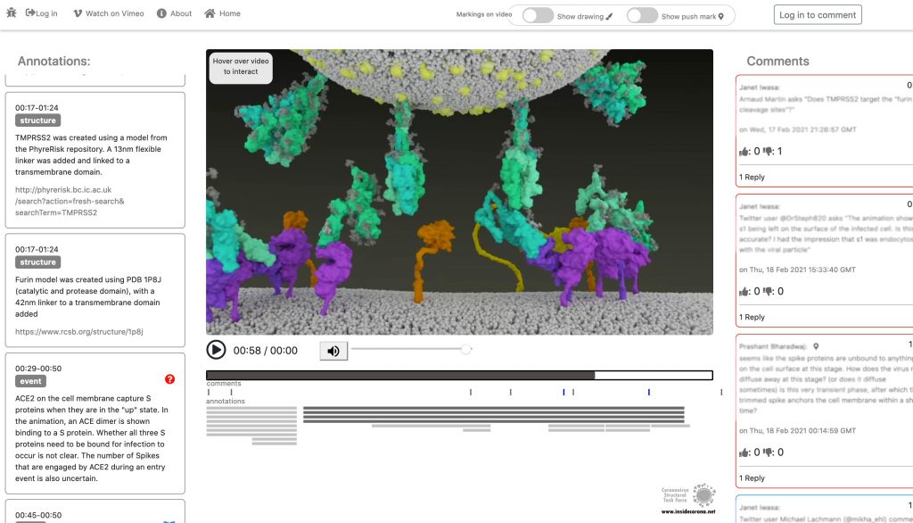
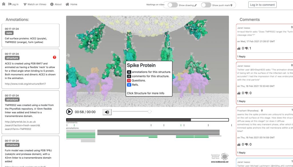
In January, this will be supplemented with a tool so that the knowledge about the SARS-CoV-2 entry mechanism can be discussed interactively by scientists all over the world. This online platform will serve as a basis for scientific discussion by providing an annotation tool. Scientific users can set a pin at any point of the video and comment their suggestions, criticism or questions about the mechanism and the structure depictions (see Fig. 1 for a prototype). Based on these annotations, the Iwasa Group will improve the animation of the entry process to provide an up-to-date detailed representation of this key process. The resulting entry animation is not only addressed to scientists, but it is also used for public outreach and education.
Even though the entry mechanism is not entirely understood yet, it could already be depicted in the fantastic animation of the Iwasa Group. There are still a lot of details and additional information to be found out about this process. From January on, the annotation tool therefore will provide the opportunity to discuss this mechanism publicly.
Thanks to the Iwasa Group for this Christmas present!

[…] Brianne – How the Body Reacts to VirusesRich – How do wombats poop cubes?Kathy – The Verse by the Side of the RoadVincent – Janet Iwasa’s SARS-CoV-2 entry animation […]
Great animation! Would like to use it for teaching purposes.
Anticipated thanks!
Hello Edmundo,
Thank you for your feedback! I am sorry to inform you that the current version of the video is under the copyright of the Iwasa Group/Thorn Group and not free for all. But there will be a release of a new version soon, which will be available for everyone. We will inform everyone about the official release on our blog, once the new version is published.
Can I share your video?
Hello Jamie,
I am sorry to inform you that the current version of the video is under the copyright of the Iwasa Group/Thorn Group and not free for all. But there will be a release of a new version soon, which will be available for everyone. We will inform everyone about the official release on our blog, once the new version is published.
[…] cool and somewhat haunting video-animation shows how SARS-CoV-2 binds and fuses to a host cell (such as one in our nasal passage) and inserts […]
[…] cool and considerably haunting video-animation exhibits how SARS-CoV-2 binds and fuses to a number cell (comparable to one in our nasal passage) […]
[…] cool and somewhat haunting video-animation shows how SARS-CoV-2 binds and fuses to a host cell (such as one in our nasal passage) and inserts […]
[…] cool and somewhat haunting video-animation shows how SARS-CoV-2 binds and fuses to a host cell (such as one in our nasal passage) and inserts […]
Can I share on YouTube, and use for teaching, with credit?
Hello Arne,
I am sorry to inform you that the current version of the video is under the copyright of the Iwasa Group/Thorn Group and not free for all. But there will be a release of a new version soon, which will be available for everyone. We will inform everyone about the official release on our blog, once the new version is published.
Great educational tool. thanks
thank you for your appreciation 🙂
What is the yellow cell surface (protein)? Is the animation intended to show the yellow cell surface protein contacting the virus particle? Is that contact significant?
What is the basis for the animation & particularly changing shape of the spike protein connecting to the cell & pulling the virus particle to the cell?
How much of this animation is actually confirmed by repeatable observation vs. surmised indirectly from observations that don't directly show changes of protein shapes?
[…] generar estrategias que controlen el contagio. Los responsables detrás del estudio fueron el Coronavirus Structural Task Force, el grupo de la investigadora Janet Iwasa, de la U. de Utah y otro grupo de expertos interesados […]
[…] 14 febrero, 2021 Vicente Noticias 0 SARS-CoV-2 Entry Animation from Iwasa Group – a little Christmas Present to the Scientific Communi… […]
[…] oben mit den Stacheln (grün). ACE2-Rezeptoren der Wirtszelle in lila. Die ganze Animation gibt es hier. (Janet Iwasa / Universität Utah und Coronavirus Structural Task […]
Brilliant