How do we know that Viruses exist?

December 22, 2020
Erik Nebelung

The SARS-CoV-2 virus has a huge negative impact on our lives, a strong contrast to the incredible small size of the virus. This elusiveness poses major challenges to our understanding and ability to fight it. In times of fake news and politicians who are abusing the pandemic for their own agenda by claiming that the virus is a hoax, there is a valid question to ask: ‘How can we be sure that the virus exists if they are even too small to be seen under the microscope?’

This question describes a scientific problem that can be traced back to the 19th century, when the true nature of viruses was a mystery even to microbiologists and doctors.

Here, we will go on a hunt for the proof of virus’s virulent existence. We will discuss the two main indications scientists look out for in matters like this, and how the scientific community solved this mystery during the past two centuries:

Indication 1: Evidence by Causality

On the first step of our hunt let us have a look at the subject: Viruses! One infamous virus is the one that is responsible for rabies. Rabies has 100% lethality after the first symptoms occur. These symptoms include fever, intense pain and a digestive tract that goes haywire. Later symptoms include hyperactivity as well as hydrophobia. This last symptom leads to the ill person developing an aversion to swallow their own saliva and feeling uncomfortable around any type of water. A terrible disease that made doctors and scientists desperately searching for the cause. But even long after bacteria had been found and identified as the origin of certain diseases (culprits for diseases are also called pathogens), no such bacteria that could be held responsible for causing rabies were ever found.

Thus the scientists of the time were confronted with a question: What exactly was responsible for diseases like rabies? Was the pathogen comparable to bacteria, or just so very alien in nature that it would not compare to anything known until then? Was it even physical in nature?
How do we know that Viruses exist? 1
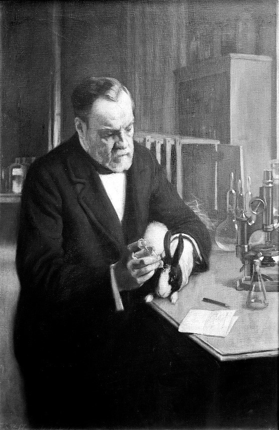
Luis Pasteur. Image by Ernest Board licensed under CC BY 4.0.

One of the persons who came one step closer to the answer of these questions was the French microbiologist Luis Pasteur, who lived in the 19th century. Previous successes with anthrax and fowl cholera established him as an icon in medicine and microbiology. But his meticulous search for the pathogen responsible for rabies was fated to be unsuccesful from day one, since none of the responsible pathogens could ever have been seen with the microscopy techniques of the time.

Given the disease’s severe nature and lethality, Pasteur tried to develop a vaccine against rabies, despite the fact that there was no hint on a pathogen’s existence except the symptoms it caused. Drawing on his experience with bacterial pathogens he eventually succeeded to develop an effective vaccine. He circumvented the need to isolate the pathogen by using infected nerve tissue to pass the disease to different mammal species like rabbits and dogs, reducing its virulence with every cycle. Just a few years later after succesfully testing it on dogs - in 1885 - the vaccine safed the life of the 9-year-old French boy Joseph Meister, making him the first person ever to be immune to rabies.

How do we know that Viruses exist? 2
Dogs, loyal companions to us humans since the stone age. And apparently not only helping us with hunting and cuddling… Image by Pixabay.

So, even when the true nature of the rabies pathogen remained a mystery, Pasteur was still able to prevent it from infecting people (and dogs), just by carefully observing the matter at hand, and learning from the way bacteria work. The fact that this technique worked implied that this pathogen was interacting with our bodies in a similar way to bacteria, proving that it must be physical in nature and hence we must be able to find it.

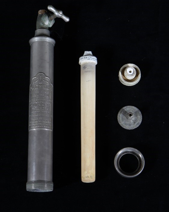
The development of the Chamberland filter, by Charles Chamberland, one of Pasteur’s assistants at the time, narrowed the physical properties of the pathogen down further. The filter basically consists of a porcelain tube with an opening on one side through which a liquid can be filtered, provided sufficient pressure.

This filter was shown to separate observable microorganisms from the liquid, including pathogenic bacteria. In 1892, Russian biologist Dimitry Iwanowski, who was investigating the mosaic disease of certain plants, filtered juice from infected leaves with the Chamberland filter and documented that the extracted juice was still infectious to healthy tobacco plants. He also observed that, unlike disease-inducing bacteria which would lose their ability to survive after a short time without nutrients, the filtrate remained infectious for many months after the filtration process. This not only added further evidence to the true nature of viruses but incidentally foreshadowed one of viruses' most annoying traits: The ability to survive without any kind of metabolism, akin to plants, animals or bacteria.
How do we know that Viruses exist? 3
Components of a Pasteur-Chamberland filter, as displayed in the Science History Institute. The white cylinder is a porcelain tube with one opening for incoming water on the top. Tightly anchored and sealed in the larger cylinder on the left, it filters the water if the incoming pressure is high enough. The filtrate leaves the setup on the other side. Image by Science history Institute licensed under CC BY-SA 3.0.

This experiment was repeated with many other diseases in the following years, including rabies. Dutch microbiologist Martinus Beijerinck first introduced the word “virus” for the filtered infectious liquid in 1898 (From Latin virus: "poison, slime, venom"). And although scientist were still uncertain if viruses were liquid or composed of particles, these experiments demonstrated that the responsible pathogens must be soluble in water and small enough to pass the Chamberland filter. This is a prime example of how scientists can demonstrate the existence of something from its effects on its environment under controlled conditions, which may even permit educated guesses about its nature.

But as they say: “Seeing is Believing”, (not "Educated Guessing is Believing") so let’s proceed our hunt for specifically visible proof with:

Indication 2: Evidence by Visualization

In the years after the discovery of the infectious liquids called “viruses” microscopy techniques improved more and more, to the point where clumps of multiple virus particles could be shown. Some scientists supposedly saw these clumped viruses without knowing it to be viruses; but the resolution and magnification abilities of light microscopes were nowhere near high enough to determine any kind of structure. There is a physical limit to the magnification a microscope can achieve. To discuss this limit, we must digress a bit and take a closer look on how waves work, and how light and matter interact:

Light can be seen as a wave. Reflection, diffraction and refraction alongside things like absorption and emission are the main interaction mechanisms between this wave and matter and they allow us to see. However, all these mechanisms depend on some level on the compared size between the waves legth and the object. Smaller objects would just be “ignored” by the wave. Since waves of any kind share some of their basic principles, a comparison with water waves can come in handy to visualize this property: Imagine boulders disrupting the waves on a beach. One could deduce the boulder's existence and make assumptions on their size by the way the waves move past these boulders and arrive on the beach. Now imagine a 20 m high super wave, which would just sweep over them and not show any sign of their existence. Similarly, electromagnetic waves ignore obstacles that are much smaller than the wavelength.​*​ This is the reason why viruses are being “ignored” by the light waves used in light microscopy. To be precise: The spectrum of visible light (which is most commonly used in light microscopy) covers wavelengths from roughly 700 nm down to ca. 400 nm. Considering the just discussed dependence of light-matter interaction on size and the way a microscope guides its light, adding the application of some math, we find the highest theoretical resolution in light microscopy to be calculated around 250 nm. Even today, however, a great expense of resources and effort must be spent to come close to this level of resolution. Most viruses are much smaller. The SARS-CoV-2 virus measures in at roughly 120 nm diameter, less than half the theoretical limit!

Now, since light microscopy cannot let us see viruses, what other way might there be to make them visible to us, on our hunt for the visible proof of viruses existence?

Enter the Electron Microscope:
How do we know that Viruses exist? 4
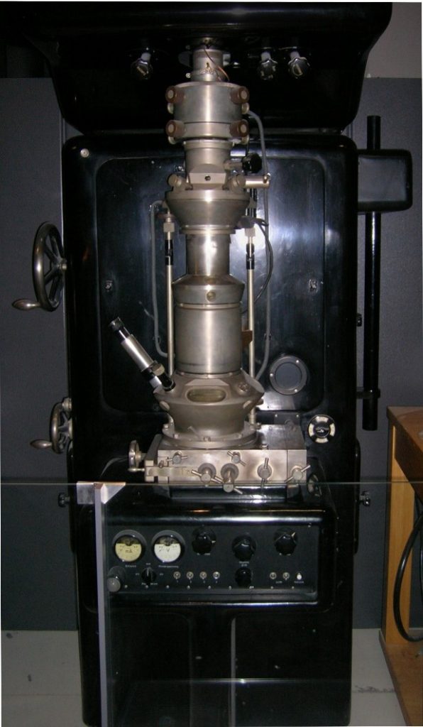
First electron microscope with resolving power higher than that of a light microscope. By Ernst Ruska, 1980. Currently displayed in the “Deutsches Museum” in Munich. Image by J. Brew licensed under DFDL.

This revolutionary device was first built by German scientists Max Knoll and Ernst Ruska in 1931. This was shortly after French physicists Lois de Broglie’s hypothesis that particles can also behave like waves went ‘viral’ in 1923. The electron microscope takes this concept and circumvents the electromagnetic wavelength problem by waving electromagnetic waves goodbye and using the wave behaviour of accelerated electrons instead. Here, the lenses which focus the light were replaced with magnetic lenses in order to focus electrons (which are charged negatively). These coil arrays produce magnetic fields that bend the pathway of the electrons. After having passed the sample, and another magnetic lens, the electrons are detected by a fluorescent screen, which translates the signals of the incoming electrons into signals we can see. Since one could theoretically accelerate the electrons to velocities very close to the speed of light - thereby shortening their wavelength more and more - the theoretical resolution limit for electron microscopes is basically unlimited.

However, relativistic effects, exponentially growing energy costs for acceleration close to the speed of light, and technical aspects of magnetic lenses limit the practical resolution limit of modern electron microscopes to some still mind blowing 0.1 - 0.2 nm.

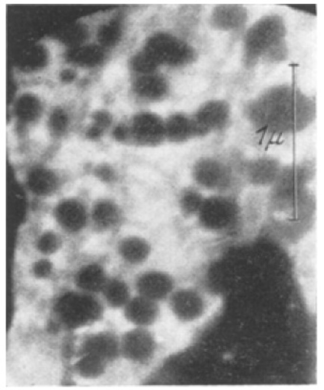
The electron microscope opened doors to deeper and smaller levels of the microcosmos for the scientific community, and as fate would have it, the brother of Ernst Ruska, Helmut Ruska, happened to be a physician and biologist. Very soon, he realized his brother’s inventions potential for microbiology, and together with their colleague Bodo von Borries they published the first microscopic picture of a virus in 1938.
How do we know that Viruses exist? 5
EM picture of the Ectromeliavirus, a virus that causes Mousepox in mice. It was discovered in 1930. This picture was taken and published in 1938 as the first micrograph of a virus. Image from "Klinische Wochenschrift 17.27 (1938): 921-925.".

What followed was a golden age for virology as the electron microscope revealed the true nature of different viruses, allowing scientists to understand how viruses operated and how to classify them.

And the new Coronavirus?

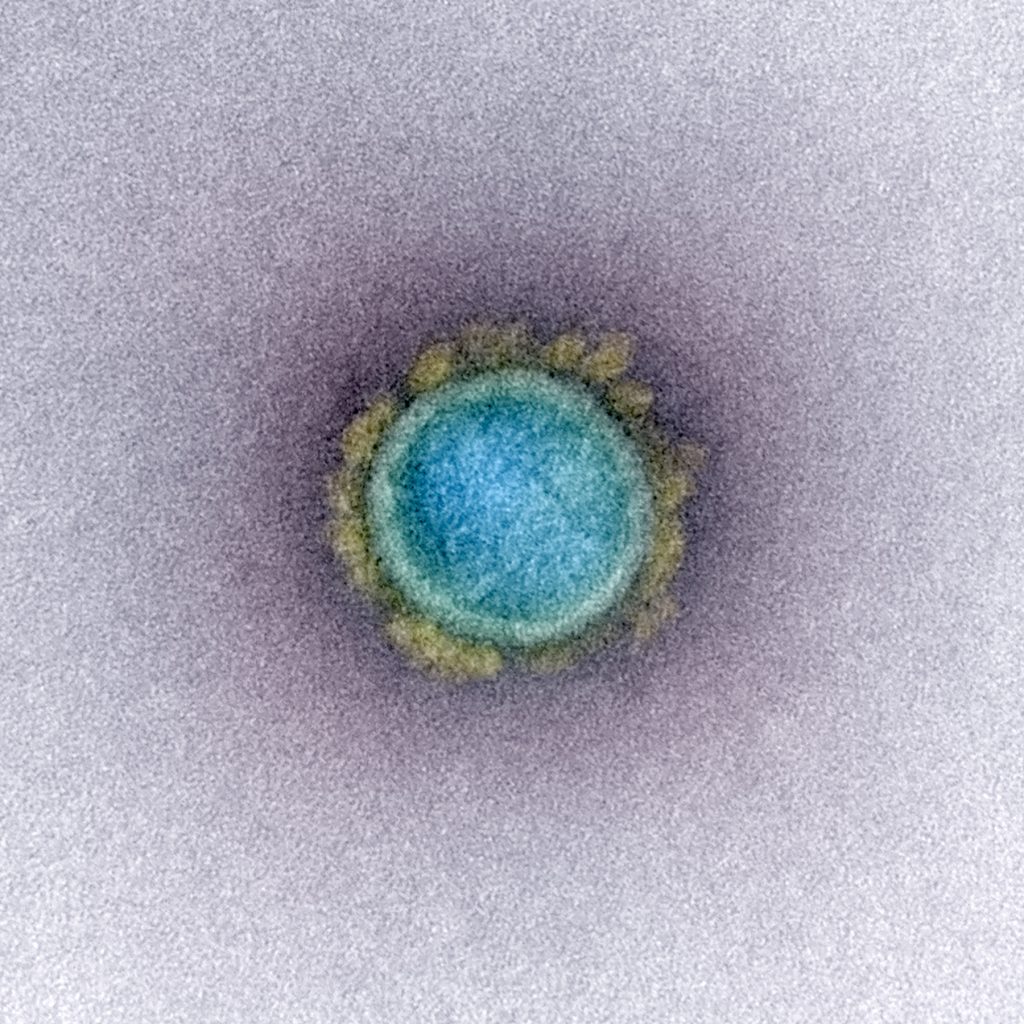
Fast forward to 2020: electron microscopy developers were not sleeping. In the 80 years that have passed since the brothers Ruska, many new methods have been developed and existing techniques were refined. And so, after the emergence of a new Coronavirus in the beginning of 2020, it was just a matter of time until EM pictures of SARS-CoV-2 were published. With this we can both see and believe SARS-CoV-2 exists!
How do we know that Viruses exist? 6
Transmission electron micrograph of a SARS-CoV-2 virus particle. Image by NIAID-RML.

Further reading and sources:

Von Borries, B., E. Ruska, and H. Ruska. "Bakterien und Virus in übermikroskopischer Aufnahme." Klinische Wochenschrift 17.27 (1938): 921-925.

Ackermann, Hans-W., and Hans-W. Ackermann. "The first phage electron micrographs." Bacteriophage 1.4 (2011): 225-227.

Knoll, Max, and Ernst Ruska. "Das elektronenmikroskop." Zeitschrift für physik 78.5-6 (1932): 318-339.

Kruger, D. H., Peter Schneck, and H. R. Gelderblom. "Helmut Ruska and the visualisation of viruses." The Lancet 355.9216 (2000): 1713-1717.

Horzinek, Marian C. "The birth of virology." Antonie van Leeuwenhoek 71.1-2 (1997): 15-20.


  1. ​*​
    Note that this analogy is not 100% accurate, but for our case this is sufficient.

Corinna the Corona Cactus

@
Corinna works as an outreach person for all plant-related business and as a mascot. She gathered previous experience in the garden center, and even though she can be a bit spiky, she likes to cuddle and lie in the sun.
More about this author

Helen Ginn

Senior Research Scientist @ Diamond Light Source, Oxfordshire, UK
Dr Helen Ginn is a senior research scientist at Diamond Light Source in the UK and a computational methods developer in structural biology. She is currently working on Representation of Protein Entities (RoPE) for structural biologists to interpret subtle conformational changes in dynamic protein systems. She has developed Vagabond for torsion angle-driven model refinement and […]
More about this author

Nick Pearce

Assistant Professor @ SciLifeLab DDLS Fellow
Nick obtained his undergraduate degree in Physics from the University of Oxford in 2012, and then his PhD in Systems Approaches to Biomedical Sciences in 2016. He moved to Utrecht in the Netherlands in 2017 to work with Piet Gros, where he obtained an EMBO long-term fellowship and worked on analysing disorder in macromolecular structures. […]
More about this author

Mathias Schmidt

Molecular Life Sciences M.Sc. Student @ Hamburg University
Mathias is currently doing his Master's degree in Molecular Life Sciences at the University of Hamburg and has been an auxiliary scientist in the Corona Structural Taskforce since March 2022. There he is working on the question of the origin of SARS-CoV-2. His undergraduate research focuses on the development of synthetic molecular mechanisms to regulate […]
More about this author

David Briggs

Principal Laboratory Research Scientist @ Francis Crick Institute in London, UK
David Briggs is a Principal Laboratory Research Scientist in the Signalling and Structural Biology lab at the Francis Crick Institute in London, UK. A crystallographer by training, his work focuses on the biophysical and structural characterisation of human extracellular proteins involved in the synapse, which have important ramifications in both psychiatric and neurodegenerative disorders. He […]
More about this author

Lisa Schmidt

Web Developer and Illustrator @ Mullana
Lisa Schmidt is a freelance illustrator who studied Multimedia and Communication (BA) in Ansbach, Germany. Her work is focused on visualising topics around science and technology. She joined the Coronavirus Structural Task Force as media designer, where she does web design, 3D rendering for scientific illustrations and outreach work.
More about this author

Philip Wehling

Nanosciences M.Sc. Student @ Institute for Nanostructure and Solid-State Physics, Hamburg University
Philip has long had an enthusiasm for biological processes which is paired with an analytical understanding of the world. After having worked for a long time as a registered nurse in various fields, he first studied mathematics and finally nanosciences. During a lecture series in preparation for a bachelor's thesis, he came into contact with […]
More about this author

Binisha Karki

Postdoctoral Research Associate @ BioNTech SE
Binisha works as a research associate at BioNTech where she works on the development of COVID-19 vaccine and cancer immunotherapies. She graduated as a Molecular Biology major from Southeastern Louisiana University in May 2019. Post-graduation she worked as a research technician in the Chodera Lab performing biophysical measurements of model protein-ligand systems for computational chemistry […]
More about this author

Binisha Karki

Wissenschaftliche Mitarbeiterin @ BioNTech SE
Binisha ist als wissenschaftliche Mitarbeiterin bei BioNTech angestellt und arbeitet an der Entwicklung von Impfstoffen gegen COVID-19 sowie Krebsimmuntherapien. Sie beendete ihr Studium der Molekularbiologie an der Southeastern Louisiana University im Mai 2019. Anschließend arbeitete sie als Forschungstechnikerin im Chodera-Lab, wo sie biophysikalische Messungen an Modellen von Protein-Liganden-Systeme für computerchemische Benchmarks durchführte.
More about this author

Hauke Hillen

Assistant Professor at the University Medical Center Göttingen & Group Leader at the MPI for Biophysical Chemistry @ University Medical Center Göttingen
Hauke ist Biochemiker und Strukturbiologe. Mit seinem Forschungsteam untersucht er mittels Röntgenkristallografie und Kryo-Elektronenmikroskopie die Struktur und Funktion von molekularen Maschinen, die für die Genexpression in eukaryotischen Zellen verantwortlich sind. Er interessiert sich dabei besonders dafür wie genetisches Material außerhalb des Zellkerns exprimiert wird, zum Beispiel in menschlichen Mitochondrien oder durch Viren im Zytoplasma.
More about this author

Richardson Lab

Richardson Lab @ Duke University, Durham, North Carolina, USA
The long-term goal of the Richardson lab is to contribute to a deeper understanding of the 3D structures of proteins and RNA, including their description, determinants, folding, evolution, and control. Their approaches include structural bioinformatics, macromolecular crystallography, molecular graphics, analysis of structures, and methods development, currently focussed on the improvement of structural accuracy. In this […]
More about this author

Holger Theymann

Agile Leadership Coach @ mehr-Freu.de GmbH
Holger keeps websites running. He makes data from scientific databases appear in nice tables. He also has an eye on keeping the sites fast, safe and reliable. His experience as a software developer, systems architect, agile project manager and coach enabled the Task Force to get the whole process well organized and he even taught […]
More about this author

Florens Fischer

Biology M.Sc. Student @ Rudolf Virchow Center, Würzburg University
Florens is studying biology (M.Sc.) and worked in the Task Force as a student assistant. He has focused on bioinformatics and supports the work on automation of scripts and structuralization of big data with machine learning. He also supported the team in other areas, such as scientific research.
More about this author

Ezika Joshua Onyeka

Public Health M.Sc. student @ Hamburg University of Applied Sciences
Joshua joined Thorn Lab as a student assistant. He is a Public Health practitioner, holds a bachelor's degree in Public Health and is currently enrolled at Hamburg University of Applied Sciences for his MPH. He has helped in implementing some vaccination programmes to improve immunisation coverage and training of immunisation frontline health workers. For the […]
More about this author

Katharina Hoffmann

Molecular Biology M.Sc. student @ Institut für Nanostruktur und Festkörperphysik, Universität Hamburg
Katharina worked as a student assistant at Thorn Lab. Normally, she studies molecular biology at the University of Hamburg. In her master's thesis, which was put on hold by Corona, she is working on the interruption of bacterial communication. Since the lockdown, she has been digging around in databases and analyzing sequences. She never thought […]
More about this author

Nicole Dörfel

Media Designer @
Nicole Dörfel ensures that we and our work are looking good! She is the illustrator, media designer and the artistic soul of the Task Force. She works her magic both in print and digitally—her focus is general media design. In the Task Force, she is mainly responsible for graphics, photo editing, design of all our […]
More about this author

Pairoh Seeliger

Administration Assistant @ Institute for Nanostructure and Solid-State Physics, Hamburg University
Pairoh Seeliger is the admin wizard of the Task Force. She takes care of media requests, handles any logistical issues that come up and makes sure our science doesn’t sound too complicated in our German outreach efforts. She self-describes as "a jack of all trades with a University education in German studies and business administration, […]
More about this author

Oliver Kippes

Biochemistry B.Sc. Student @ Rudolf Virchow Center, Würzburg University
Oli is studying biochemistry (B.Sc) and has completed a training as an IT specialist prior to his studies. With the combined knowledge of his studies and training, he helps maintaining the structural database, programs applications for it and supports the team in literature research. In spite of his study, structural biology was still a new […]
More about this author

Luise Kandler

Biochemistry B.Sc. Student @ Rudolf-Virchow Center, Würzburg University
Luise is a B.Sc. student in biochemistry at the University of Würzburg and joined the Task Force during the first Corona lockdown. She did her bachelor's thesis with the Thorn Lab, where she learned programming with Python and worked on the implementation of a GUI for our machine learning tool HARUSPEX in Coot. In the […]
More about this author

Ferdinand Kirsten

Biochemistry B.Sc. Student @ Rudolf Virchow Center, Würzburg University
Ferdinand did his bachelor's thesis at Thorn Lab on solvent exchange and interactions in macromolecular crystallography. Still new to the world of crystallography and structural refinement, he tries to help wherever he can, with a main focus on literature and genome research as well as structural refinement with Coot. Even if he's more of the […]
More about this author

Kristopher Nolte

Biochemistry B.Sc. Student @ Rudolf-Virchow Center, Würzburg University
Kristopher joined Thorn Lab as part of his bachelor thesis. In this thesis he refined aspects of the diagnostic tool for graphical X-Ray data analysis (AUSPEX) with the help of machine learning. But since the corona crisis halted all our lives, he contributes to the Task Force by using his knowledge of bioinformatics and programming […]
More about this author

Erik Nebelung

Nanoscience M.Sc. Student @ Institute for Nanostructure and Solid-State Physics, Hamburg University
Erik is studying nanoscience with a focus on biochemical methods and applications. From August 2020 till January 2021 he pursued his studies at the iNano institute in Aarhus, before starting his master's thesis back in Hamburg. He had his first taste of protein crystallization during his bachelor's thesis work and this sparked his interest in […]
More about this author

Toyin Akinselure

Nanoscience M.Sc. Student @ Institute for Nanostructure and Solid-State Physics, Hamburg University
Toyin ist a microbiologist and presently an M.Sc. student in nanoscience with a focus on nanobiology and nanochemistry. She is interested in scientific research especially in protein chemistry and drug discovery. In the previous autumn and winter, she interned with two research projects, one in drug discovery and the other in protein structure. She found […]
More about this author

Lea von Soosten

Physics M.Sc. Student @ Institute for Nanostructure and Solid-State Physics, Hamburg University
Lea is a M.Sc. physics student with a great interest in everything related to biology. Even though she comes from a different field, she joined the team to expand her knowledge in biochemistry and help the Task Force with a main focus on literature research. Also, she loves drawing!
More about this author

Sabrina Stäb

Biotechnology M.Sc. Student @ Institute for Nanostructure and Solid-State Physics, Hamburg University
Sabrina is studying biochemistry (M.Sc.) and works as a research assistant for the Thorn Lab and the CSTF. During her bachelor thesis on "Crystallization and Structure Solution of High-Quality Structures for MAD Experiments", she was able to gain a lot of experience in the field of crystallography and now brings this experience to the project. […]
More about this author

Alexander Matthew Payne

Chemical Biology Ph.D. Student @ Chodera Lab, Memorial Sloan Kettering Center for Cancer Research, New York, U.S.
Alex is a Ph.D. student interested in understanding how proteins move! He has recently joined the labs of John Chodera and Richard Hite to work on a joint project involving molecular dynamics and Cryo-EM. His goal is to generate conformational ensembles from Cryo-EM data and simulate the ensemble using massive scale molecular dynamics via Folding@Home. […]
More about this author

Maximilian Edich

Bioinformatics Ph.D. Student @ Institute for Nanostructure and Solid-State Physics, Hamburg University
Max studied bioinformatics and genome research in Bielefeld and joined the CSTF as a Ph.D. student in 2021. Previously, his focus was on molecular modeling. Now, he works on the so-called R-factor gap. He already learned what it is like to be part of a young, scientific team as a member of the iGEM contest […]
More about this author

Agnel Praveen Joseph

Computational Scientist @ Science and Technology Facilities Council, UK
Dr. Agnel Praveen works as a computational scientist in the CCP-EM team at the Science and Technology Facilities Council, UK. He is interested in approaches to interpret and validate maps and atomic models derived from Cryo-EM data and looks also into computational methods for the interpretation of Cryo-ET data. In collaboration with five other sites […]
More about this author

Dale Tronrud

Research Scientist @
Dale Tronrud has both solved protein crystal structures and developed methods and software for the optimization of macromolecular models against X-ray data and known chemical structural information. He has had a long-standing interest in enzyme:inhibitor complexes and photosynthetic proteins, focusing on the Fenna-Matthews-Olson protein. In addition, he has also been involved in the validation and […]
More about this author

Sam Horrell

Beamline Scientist @ Diamond Light Source, Oxfordshire, UK
Sam is a structural biologist working on method development around structural biology at Diamond Light Source, in particular for ways of better understanding how enzymes function through the production of structural movies. Sam is working through deposited structures related to SARS-CoV and SARS-CoV-2 with a view to providing the most accurate protein structures possible for […]
More about this author

Cameron Fyfe

Postdoctoral Research Associate @ Micalis Institute, INRAE, Paris, France
Cameron is a structural biologist who has worked extensively on proteins from microorganisms. With many years of experience in the pharmaceutical industry and in structural biology research, he joined the Task Force to contribute his skills to improve existing models for drug development. He is currently researching Radical SAM enzymes at INRAE. When not in […]
More about this author

Tristan Croll

Postdoctoral Research Associate @ Cambridge Institute for Medical Research, University of Cambridge
Tristan is a specialist in the modelling of atomic structures into low-resolution crystallographic and cryo-EM density, and developer of the model-building package ISOLDE. His focus in the project is on correcting the various errors in geometry and/or chemical identity that tend to occur in less well-resolved regions, with the overall aim of bringing the standards […]
More about this author

Gianluca Santoni

Serial Crystallography Data Scientist @ European Synchrotron Radiation Facility, Grenoble, France
Gianluca is an expert in protein crystallography data collection and analysis. After a PhD in structure-based drug design, he has worked as a postdoc on the beamline ID23-1 at the European Synchrotron Radiation Facility (ESRF) and has developed the SSX data analysis software ccCluster. His current interests are the optimization of data collection strategies for […]
More about this author

Yunyun Gao

Postdoctoral Research Associate in the AUSPEX Project @ Institute for Nanostructure and Solid-State Physics, Hamburg University
Yunyun is a method developer for strategies of analysing data from biomacromolecules. Before joining the Thorn group, he had been working on SAXS/WAXS of polymers and proteins. He is interested in improving objectivity and reliability of data analysis. Yunyun is currently extending the functionality of AUSPEX. He is the repository manager and AUSPEX handler for […]
More about this author

Johannes Kaub

Scientific Coordinator @ Institute for Nanostructure and Solid-State Physics, Hamburg University
Johannes Kaub studied chemistry at RWTH Aachen, with a focus on solid-state physical chemistry, before serving as a scientific employee at the Max Planck Instiute for the Structure and Dynamics of Matter. He supports the Coronavirus Structural Task Force as a scientific coordinator with his organizing ability and his talent for solving problems. Other than […]
More about this author

Andrea Thorn

Group Leader @ Institute for Nanostructure and Solid-State Physics, Hamburg University
Andrea is a specialist for crystallography and Cryo-EM structure solution, having contributed to programs like SHELX, ANODE and (a little bit) to PHASER in the past. Her group develops the diffraction diagnostics tool AUSPEX, a neural network for secondary structure annotation of Cryo-EM maps (HARUSPEX) and enables other scientists to solve problem structures. Andrea is […]
More about this author

6 comments on “How do we know that Viruses exist?”

  1. those arent pictures of viruses those are the proteins that our body produces to fight the infection aka our immune response

    1. What exactly are you talking about? There are cells (B lymphocytes) that produce antibodies yes, and after recieving an mRNA based vaccine, some bodycells would produce the SARS-CoV-2 spike protein. Both would look much different though compared to the picture. Human cells would be much much larger in comparison to the proteins they would produce. And Antibodies are even smaller than the spikes. Further: When this EM picture was made, no mRNA vaccines even existed yet. This is a virus and its spike proteins.

  2. Your article ended far too soon. You show us a picture and conclude that it is a virus. That is not proof, it is just a picture.

    Were you able to isolate and characterize the virus? What technique did you use to isolate it for the TEM picture?. These are important questions. Were you able to meet Koch's Postulates for your isolation procedure?

    If your intent is to be credible then you need to also publish your methodology, extraction, purification, and isolation techniques. The process of science requires you to follow these protocols, not to jump to your conclusion.

    1. Dear Terry Maurice, this is a blogpost aimed to explain some scientific background of virology for everyone. This is NOT a scientific work/publication for a scientific journal, in which methodology etc. are - as you rightly state - obligatory and important parts of the articles. The picture I used is from the "The National Institute of Allergy and Infectious Diseases". A similar picture of the virus, embedded in a scientific article by a Japanese research group, can be found here: https://www.pnas.org/doi/epdf/10.1073/pnas.2002589117 Sadly, scientific articles tend to be hard to comprehend even for scientists. This is because of the heavy use of technical terms which is necessary to keep the articles short.

      Further: I recommend this article about Koch's Postulates and why they are partially outdated, and how to assess them in the context of SARS-CoV-2: https://www.reuters.com/article/factcheck-koch-idUSL2N2L23F1 .

      Kind Regards

Leave a Reply

Your email address will not be published. Required fields are marked *

cross